AI reveals sex-related differences in brain microstructure

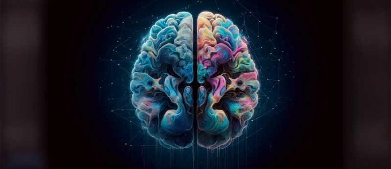
A recent study led by researchers at NYU Langone Health (NY, US) has demonstrated the power of AI combined with diffusion MRI (dMRI) to uncover sex-related differences in human brain microstructure.
Using a multi-model approach, the study analyzed thousands of dMRI brain scans from 471 men and 560 women, revealing that the brains of men and women are organized differently at a cellular level, particularly in white matter, which facilitates communication between brain regions.
The importance of understanding brain organization
Men and women experience conditions such as multiple sclerosis, autism spectrum disorder, and migraines at different rates and with varying symptoms. Understanding how biological sex impacts brain organization is crucial for improving diagnostics and treatments. Previous studies focused on brain size, shape, and weight but lacked detailed insight into cellular-level organization. This study fills that gap by using AI to detect structural patterns in white matter that are invisible to the human eye.
AI models employed in the study
The researchers employed three different AI models—2D convolutional neural networks (CNNs), 3D convolutional CNNs, and Vision Transformers (ViTs) —to analyze the dMRI data. Here’s a breakdown of how each was used:
2D CNN
Based on a ResNet18 architecture, this model processes 2D slices of the 3D brain volume. Specifically, the researchers extracted features from every three consecutive slices of the brain images and combined them for analysis. Features from all subvolumes were concatenated and fed into a linear prediction layer to classify the sex of the subject.
3D CNN
Based on a 3D ResNet10 architecture, this model processes the entire 3D volume directly to establish local and global spatial relationships between structures in the data. Features were pooled and fed into a fully connected layer to make the final sex classification.
Vision Transformers
The ViT is originally designed for 2D image recognition but was adapted for 3D input in this study. The 3D brain volume was divided into non-overlapping 3D patches. Each patch represents a small cube of the brain volume, containing detailed local information. Each patch was encoded with its location in the brain. These patches were processed by the ViT, which used self-attention to capture both detailed and overall patterns. A classification token (think of this like a summary note of a long document) collated this information to determine the brain scan’s sex.
Results and key findings
Together, these models could accurately distinguish between male and female brains, with an accuracy of 92% to 98%.
To identify specific white matter regions contributing to sex classification, the researchers used occlusion analysis. This involved systematically occluding (masking) different regions of the brain and observing changes in the model’s classification performance. The analysis revealed key white matter regions, such as the corpus callosum and middle cerebellar peduncle, which showed significant sex-related differences.
“Our findings provide a clearer picture of how a living, human brain is structured, which may in turn offer new insight into how many psychiatric and neurological disorders develop and why they can present differently in men and women,” said study senior author Yvonne Lui, MD, a neuroradiologist at NYU Grossman School of Medicine.
Moving beyond traditional research methods
The study’s approach differed from previous research, which often relied on subjective statistical analyses of specific brain regions. By using machine learning to analyze entire groups of images, the study avoided human biases. The AI models, trained with brain scans from healthy individuals labeled by biological sex, learned to distinguish sex-related differences on their own, focusing on features like the movement and direction of water through brain tissue.
In the future, the research team plans to explore how sex-related brain structure differences develop over time, considering environmental, hormonal, and social factors.


