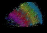
EMDiffuse: AI-Powered Enhancement for Electron Microscopy

Researchers have developed EMDiffuse, a suite of AI algorithms designed to enhance electron microscopy (EM) imaging. Published recently in Nature Communications, this study explores how these algorithms might improve the resolution and speed of EM techniques, potentially expanding our ability to visualize cellular structures.
Electron Microscopy: Power and Limitations
Instead of using photons (light particles) as in traditional light microscopes, EM uses electrons.
Electrons have much shorter wavelengths than visible light, allowing for higher resolution imaging, meaning finer details can be observed. This makes it extremely powerful for visualizing nanoscale biological structures such as DNA, viruses and cellular organelles.
Volume EM (vEM) extends this capability to three dimensions, allowing visualization of larger structures like whole cells or tissues.
While EM is incredibly useful, it has some notable limitations:
- Trade-offs between imaging speed, quality, and sample size
- Difficulty in achieving high-resolution in all dimensions
- Noise interference, particularly when looking “deep” into a sample
While previous AI approaches attempted to address these issues, they often resulted in smoothed images with reduced resolution.
EMDiffuse: A New Approach
EMDiffuse, developed by Chixiang Lu and colleagues, uses diffusion-based AI models to overcome these limitations.
Diffusion models work by gradually adding noise to an image and then training the AI to reverse this process. This approach allows the AI to learn how to reconstruct high-quality images from noisy ones, preserving important structural details while removing unwanted noise.
The EMDiffuse suite includes several components:
- EMDiffuse-n: For denoising 2D images
- EMDiffuse-r: For super-resolution tasks, improving image clarity
- vEMDiffuse-i: For creating high-quality 3D images using pre-existing high-resolution 3D data for training
- vEMDiffuse-a: For creating high-quality 3D images using only lower-quality 3D data for training
Performance and Applications
EMDiffuse significantly improves both image quality and acquisition speed. For instance, EMDiffuse-r doubled image resolution while increasing EM imaging speed by 36 times in their setup.
The researchers demonstrated EMDiffuse’s effectiveness in:
- Clearly show the shapes of cell organelles in 3D
- Help trace the paths of nerve cells in brain tissue
- Improve images much faster than current methods
Importantly, EMDiffuse is highly adaptable, requiring minimal fine-tuning for new types of biological samples.
Implications
This advancement opens new possibilities scientists studying how cells work, how diseases affect cells, or how drugs interact with cells. For example, it might let researchers see larger pieces of tissue in detail, or observe changes in cells more quickly.
While EMDiffuse shows promise, it’s not perfect. It works best when the original images are of a certain quality.
This requirement means that while EMDiffuse can significantly enhance images, it’s not a magic solution for poor-quality data. Researchers still need access to relatively high-quality electron microscopes to fully benefit from EMDiffuse’s capabilities.
Future Directions
The researchers note that improving EMDiffuse’s performance on lower-resolution data is an area for future development. This could potentially make the technology more accessible to a wider range of researchers and institutions.
In conclusion, EMDiffuse represents a significant leap forward in AI-enhanced EM, offering researchers powerful new tools to explore the intricate world of cellular structures. This technology has the potential to dramatically accelerate biomedical research and drug discovery processes.
